Exhibition shows animals inside out
Read this article for free:
or
Already have an account? Log in here »
To continue reading, please subscribe:
Digital Subscription
One year of digital access for only $75*
- Enjoy unlimited reading on winnipegfreepress.com
- Read the E-Edition, our digital replica newspaper
- Access News Break, our award-winning app
- Play interactive puzzles
*Billed as $5.77 plus GST every four weeks. After 52 weeks, price increases to the regular rate of $19.95 plus GST every four weeks. Offer available to new and qualified returning subscribers only. Cancel any time.
Monthly Digital Subscription
$4.99/week*
- Enjoy unlimited reading on winnipegfreepress.com
- Read the E-Edition, our digital replica newspaper
- Access News Break, our award-winning app
- Play interactive puzzles
*Billed as $19.95 plus GST every four weeks. Cancel any time.
To continue reading, please subscribe:
Add Free Press access to your Brandon Sun subscription for only an additional
$1 for the first 4 weeks*
- Enjoy unlimited reading on winnipegfreepress.com
- Read the E-Edition, our digital replica newspaper
- Access News Break, our award-winning app
- Play interactive puzzles
*Your next Brandon Sun subscription payment will increase by $1.00 and you will be charged $17.95 plus GST for four weeks. After four weeks, your payment will increase to $24.95 plus GST every four weeks.
Read unlimited articles for free today:
or
Already have an account? Log in here »
Hey there, time traveller!
This article was published 18/04/2019 (2638 days ago), so information in it may no longer be current.
Nobody is talking about the elephant in the room.
To be fair, it’s merely a cross-section of an elephant, and people are busier talking about the giraffe and the ostrich and the camels.
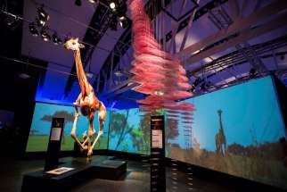
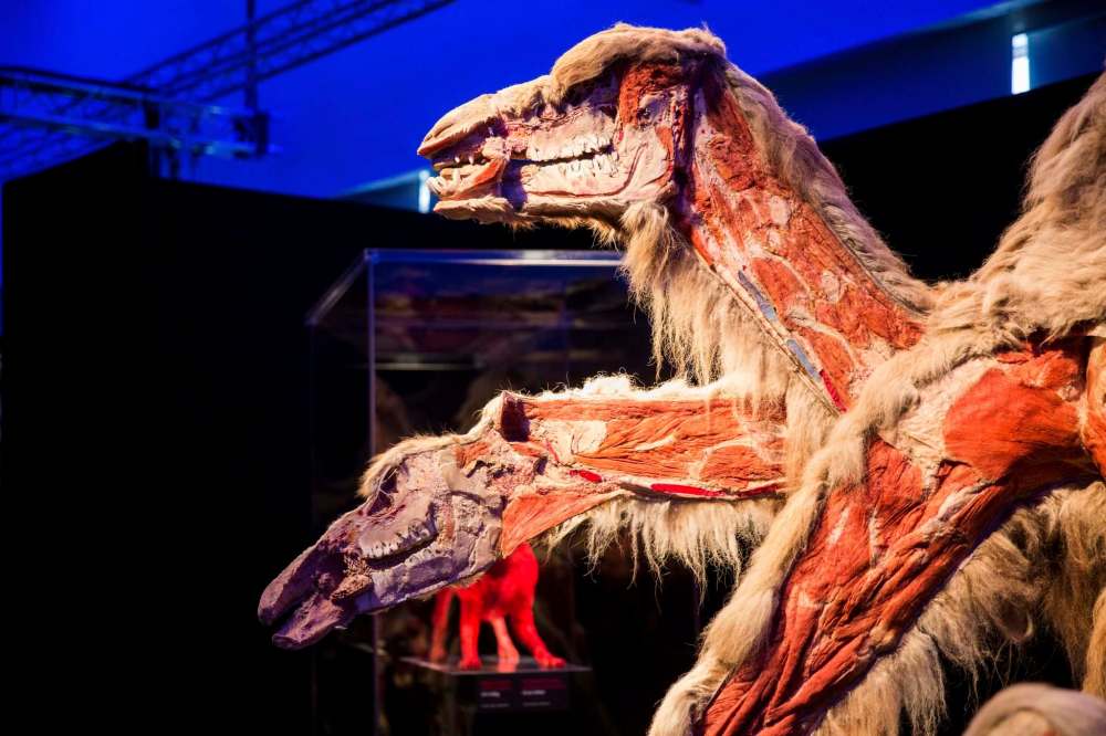
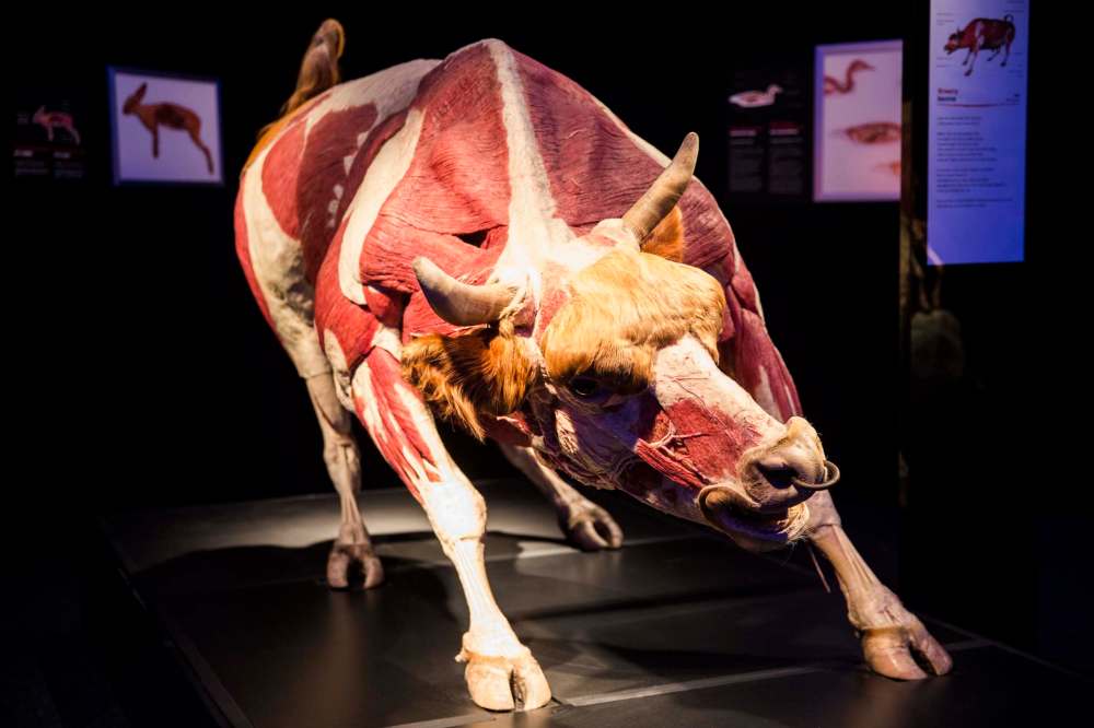
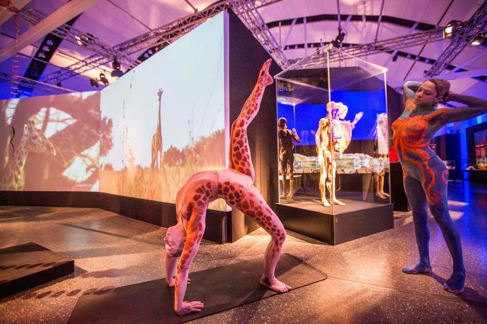
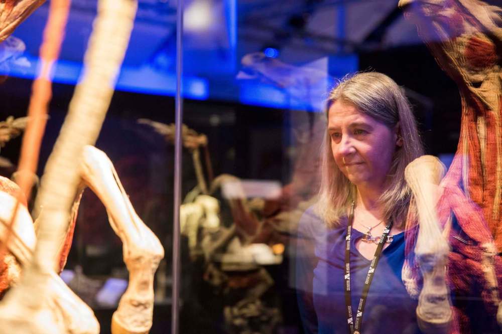
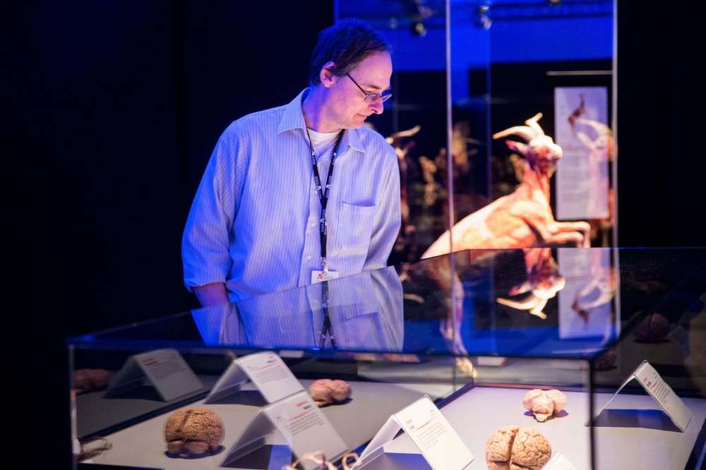
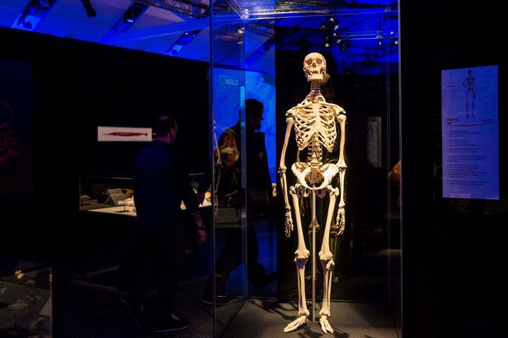
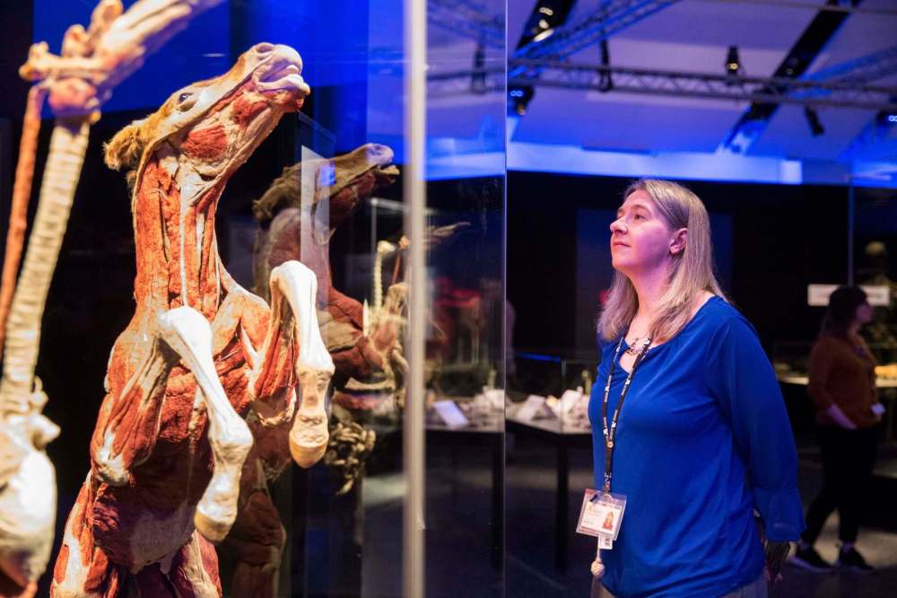
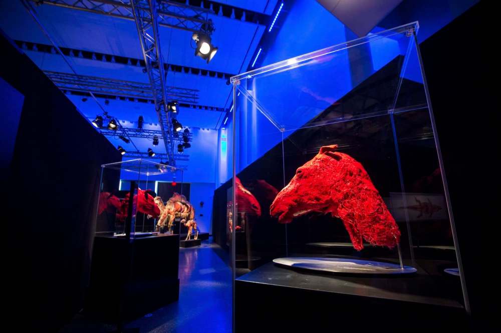
These creatures, posed in lifelike postures but with their musculature, tendons and ligaments dramatically and skinlessly exposed, are part of the Body Worlds: Animal Inside Out exhibition on at the Manitoba Museum.

Preserved via the process of plastination, in which the water in the tissues is replaced by a polymer such as epoxy resin or silicon rubber, at first glance, the flayed appearance of the animals is shocking, but closer examination reveals a curious kind of beauty. Whether it’s the heavily muscled shoulders of a bull, the stiff gills and terrifying maw of a shark or the ruminant stomach of a snow camel, it’s a glimpse into the anatomy and biology of animals we might never see hide nor hair of, let alone hideless and hairless with their inner workings on display.
“You gain a different perspective on nature and these intricate designs, and appreciate life even more,” says curator Dr. Angelina Whalley of the animals, organs and bodily systems on display in the exhibition, which runs to Sept. 2. “This is my sincere goal with Animals Inside Out, that people leave the exhibition with an idea of respect and appreciation of all these animals, and life in general.”
Plastination process
The inventor of plastination, Dr. Gunther von Hagens, came to his unusual vocation in a roundabout way.
The inventor of plastination, Dr. Gunther von Hagens, came to his unusual vocation in a roundabout way.
Born in East Germany, before the fall of the Berlin Wall, “he wanted to flee, to escape,” says his wife, Dr. Angelina Whalley. “He was caught and put in political prison for more than two years.”
Later, as a young physician, he dreamed of a life of freedom in the United States. Von Hagens decided his ticket out of East Germany was to get certification from the Educational Commission for Foreign Medical Graduates, which assesses whether medical grads are ready to enter residence programs in the U.S. He needed time to prepare and study, but he worked so many night shifts and weekends at the hospital, he couldn’t find the time.
“He applied for a job in the anatomy lab,” Whalley says. “It was the opportunity to earn money for daily life and to prepare for the U.S. During his research work, which was very boring to him, he came up with the idea for plastination and he was so caught with it. He thought, ‘This will revolutionize teaching anatomy.’“
The process is under constant refinement, but basically works like this:
• Decomposition is halted by embalming the cadaver or carcass in formaldehyde.
• Specimens are dissected with forceps and scalpels
• Frozen bodily fluids are replaced by acetone in a cold acetone bath
• Soluble fat molecules are replaced by acetone in a warm acetone bath
• In a vacuum, acetone is extracted and gradually replaced with plastic
• Each structure is brought into the proper position
• After gas curing, the posed specimen is infused with durable silicon rubber
— Source: Body Worlds
Whalley is the director of the Institute of Plastination in Heidelberg, Germany, and the conceptual planner and creative designer of Body Worlds, the travelling expo of dissected human bodies, seen by more than 47 million people worldwide, which relies on the preservation process invented by her husband, Dr. Gunther von Hagens, in 1977. (Body Worlds’ human cadavers have been freely donated; the company has not been dogged by the same controversy as Bodies: The Exhibition, which visited Winnipeg in 2010. Protesters alleged the cadavers were the bodies of political prisoners and Falun Gong followers who had been imprisoned in China.)
Most of the animals in Animal Inside Out were donated by zoos and game parks all over the world, where they were declared to have died of natural causes. In a process that can take up to two years, they have been plastinated, some in whole-animal form, others dissected and rendered into thin translucent slides to show a cross-section of the inner systems and organs. One of the giraffes, displayed as an expanded array of its cross-sectioned slices, took 20,000 hours to dissect.
Though there are about 400 plastination labs in the world, the institute is uniquely qualified to handle large animals, which must be submerged whole into an acetone bath as part of the process.

They also require quicker treatment to prevent purification than human bodies (which may be displayed as part of Body Worlds exhibits or used as specimens by medical schools or anatomy labs).
“A human cadaver comes to our institute typically two to three days after the person has passed away,” Whalley says. “Animals, we appreciate to have them earlier because when they are vegetarians, they typically have a different bowel and can develop a lot of gas inside.”
After they’ve been preserved, positioning the animals and shaping them into posture takes another six months or so.
“For exhibition purposes, we like a very natural, more or less moving pose,” she says. “When we had our very first exhibition in Japan, there was only one sincere complaint. People said, ‘They are interesting but they are also looking a little frightening; they look so dead.’ So we understood that a specimen for public display needs to have an additional quality other than just a scientific expression.”
Whalley has curated the exhibition to focus on several different areas: skeletal foundations; muscles, tendons and ligaments; the nervous system; reproduction; breathing and eating; and wildlife conservation and preservation. She has included examples of human anatomy for comparison and by highlighting the similarities and unique qualities of each species, hopes to inspire a sense of appreciation for the natural world.
“The animal kingdom is very diverse, so it’s hard to focus on one thing,” she says.
“So I decided to refer to bodily functions and that helped me to create a bridge to the human anatomy, so people are aware that we function the same way with the same structures; it’s all the same.”
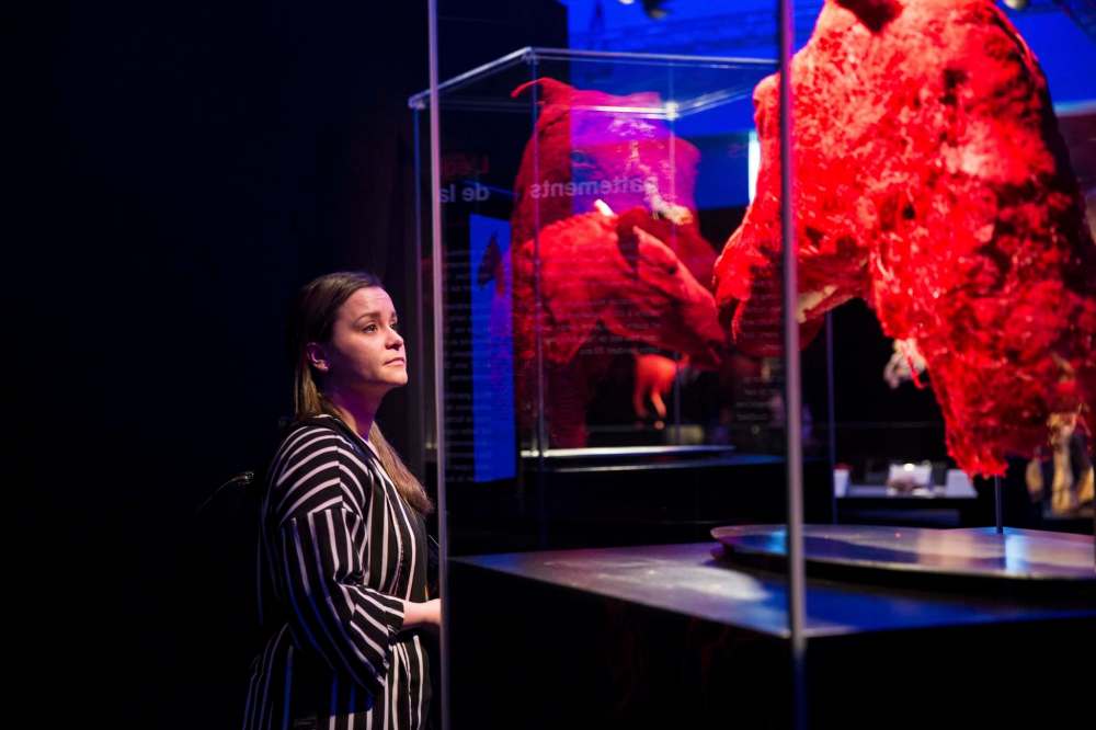
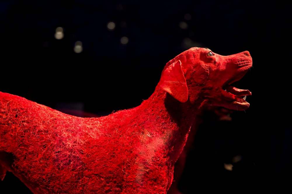
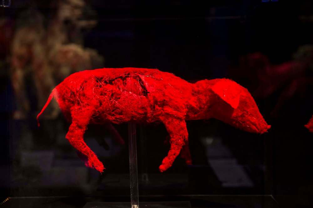
Some of the most striking exhibits are the structures showing the dense, intricate web of the circulatory system of a rabbit or a dog. These are some of Whalley’s favourites, both for their delicate esthetic appeal and for their illustration of the strides Body Worlds is taking in improving plastination technology.
“We inject a red polymer into the arteries,” Whalley says, explaining that a large part of the institute’s work revolves around polymer-chemistry research. “When the polymer cures, it takes over the shape of the system. Then we can dissolve the tissue away by means of enzymes or bacteria. We would never be able to dissect manually in that much detail. What you get is a cast of the arterial system, and you need a very particular polymer (to achieve it).”
She is aware that some potential viewers find the idea of the exhibition distasteful or even frightening.
“I often hear that and I can understand that,” she says. “But even when people come with hesitation, it really, for most people, is breathtaking. They often say, ‘It is completely different from what I expected; it is so beautiful. I’m glad I came because it’s really an eye-opener.’
“It offers a view into something you believe you know, but you really know very little about.”
jill.wilson@freepress.mb.ca
Twitter: @dedaumier


Jill Wilson started working at the Free Press in 2003 as a copy editor for the entertainment section.

Our newsroom depends on a growing audience of readers to power our journalism. If you are not a paid reader, please consider becoming a subscriber.
Our newsroom depends on its audience of readers to power our journalism. Thank you for your support.